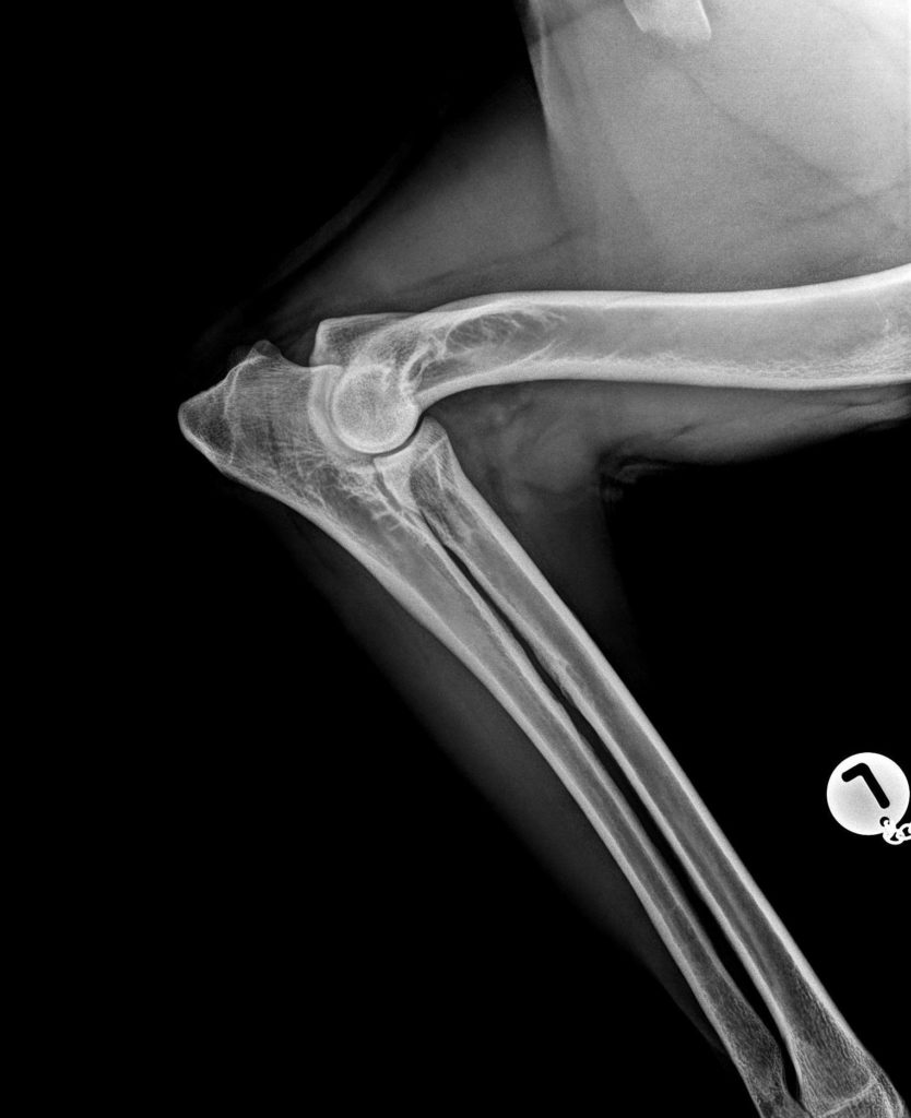
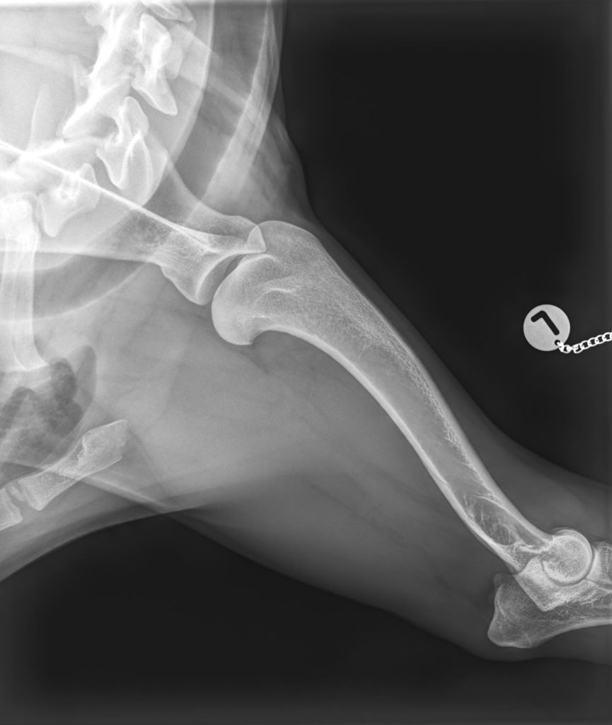
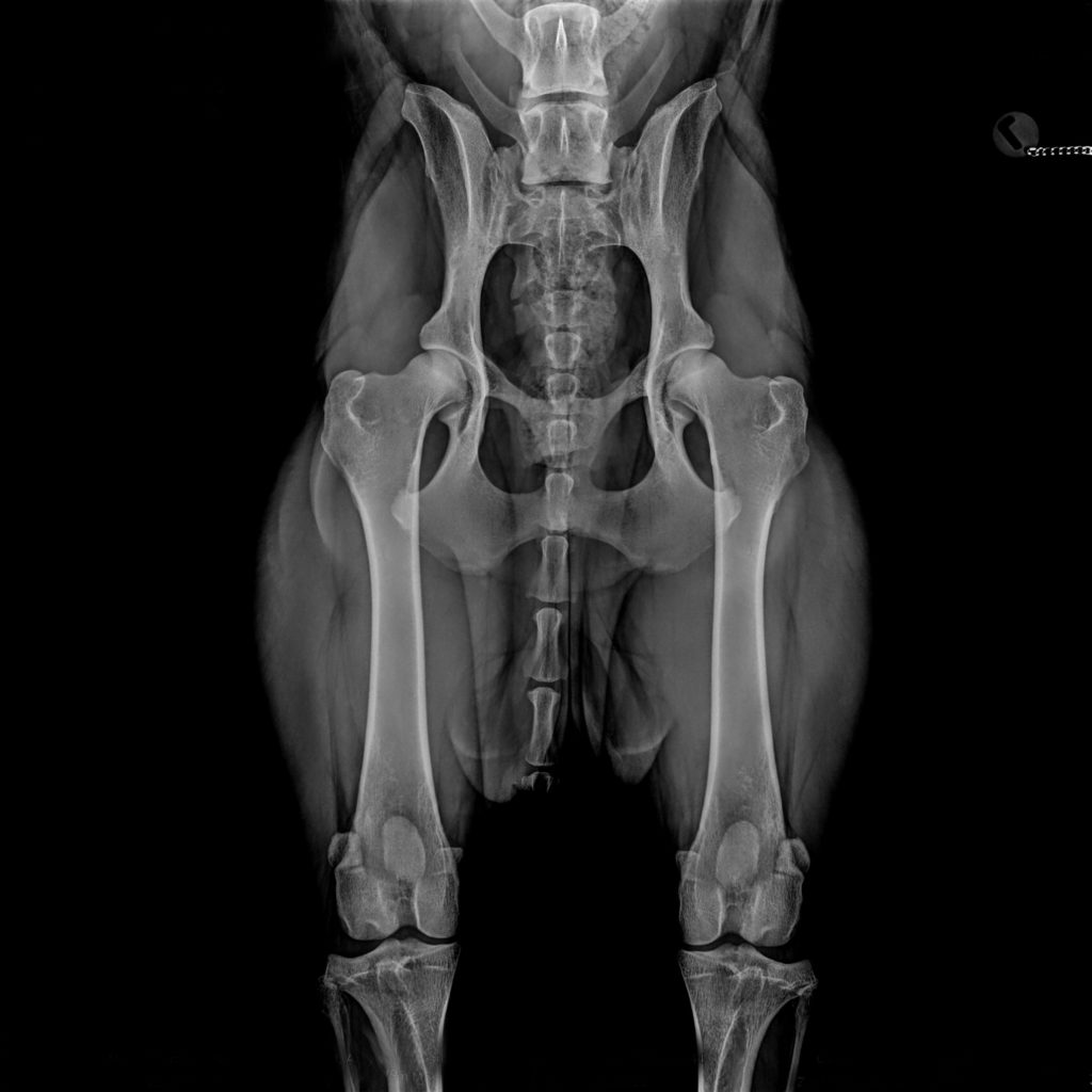
Official HD, ED, OCD screening
As part of the breeding examinations for the German Longhaired Pointer (Deutsch Langhaar), we take and evaluate official HD (Hip Dysplasia), ED (Elbow Dysplasia), and OCD (Osteochondrosis Dissecans) X-rays. We also evaluate images taken elsewhere, provided they comply with the official guidelines. In the following text, you will find detailed information regarding the registration form, radiographic techniques, and explanatory images.